When Your Liver Says No: Understanding Obstructive Jaundice
Jaundice, the yellowing of the skin and whites of the eyes, often signals an underlying issue with bilirubin, a yellow pigment produced by the breakdown of red blood cells. In obstructive jaundice, the normal flow of bilirubin is blocked, causing it to accumulate in the bloodstream and leak into tissues. This blog dives deep into obstructive jaundice, exploring its types, causes, how it develops, diagnosis methods, treatment options, prevention strategies, and concluding with key takeaways.
A Block in the Flow: Types of Obstructive Jaundice
Obstructive jaundice occurs when there is a blockage in the bile ducts that carry bile from the liver to the gallbladder and small intestine. This blockage prevents bile from flowing normally, leading to the buildup of bilirubin in the blood and causing jaundice. Obstructive jaundice can be classified based on the location of the obstruction:
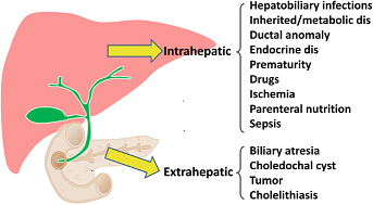
1. Intrahepatic Obstruction
Intrahepatic obstructive jaundice occurs within the liver. This type of obstruction is typically due to diseases or conditions that affect the bile ducts inside the liver.
Causes of Intrahepatic Obstruction:
- Hepatitis: Inflammation of the liver caused by viral infections, alcohol, or drugs.
- Cirrhosis: Scarring of the liver tissue that disrupts normal liver function and bile flow.
- Primary Biliary Cholangitis (PBC): An autoimmune disease that gradually destroys the bile ducts within the liver.
- Primary Sclerosing Cholangitis (PSC): Chronic inflammation and scarring of the bile ducts inside and outside the liver.
- Liver Tumors: Benign or malignant tumors within the liver that compress bile ducts.
2. Extrahepatic Obstruction
Extrahepatic obstructive jaundice occurs outside the liver. This type of obstruction is typically due to physical blockages in the bile ducts located outside the liver.
Causes of Extrahepatic Obstruction:
- Gallstones: The most common cause; stones can obstruct the common bile duct.
- Biliary Strictures: Narrowing of the bile ducts due to inflammation, surgical injury, or trauma.
- Pancreatic Tumors: Tumors, especially in the head of the pancreas, can compress the bile duct.
- Cholangiocarcinoma: Cancer of the bile ducts, which can block bile flow.
- Ampullary Cancer: Cancer at the junction where the bile duct and pancreatic duct meet the small intestine.
- Choledochal Cysts: Congenital cysts that cause dilation and obstruction of the bile ducts.
- Parasites: Infections such as liver flukes can obstruct bile ducts.
3. Mixed-Type Obstruction
In some cases, obstructive jaundice may involve both intrahepatic and extrahepatic components. This can occur in conditions that cause extensive damage to the bile ducts both inside and outside the liver.
Causes of Mixed-Type Obstruction:
- Advanced Primary Sclerosing Cholangitis (PSC): Can involve scarring and narrowing of bile ducts both inside and outside the liver.
- Metastatic Cancer: Cancers from other organs can spread to both intrahepatic and extrahepatic bile ducts, causing blockages in multiple locations.
Pathogenesis of Obstructive Jaundice
The pathogenesis involves the following steps:
-
Bile Flow Obstruction:
- Obstruction can occur due to a mechanical blockage (e.g., stones, tumors) or narrowing of the bile ducts.
-
Bile Accumulation:
- Bile, which contains bilirubin, accumulates in the liver and spills over into the bloodstream, leading to hyperbilirubinemia.
-
Tissue Deposition:
- Elevated bilirubin levels result in its deposition in tissues, causing the yellow discoloration of the skin and eyes (jaundice).
-
Secondary Effects:
- Bile stasis can lead to infections (cholangitis) and liver damage (biliary cirrhosis) if left untreated.
Clinical Features of Obstructive Jaundice
The clinical features can be divided into general symptoms, signs related to the biliary system, and systemic manifestations.
1. General Symptoms
Jaundice:
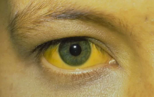
- Yellowing of the Skin and Eyes: The hallmark symptom of obstructive jaundice. It occurs due to elevated levels of bilirubin in the blood, which deposits in the skin and sclera (the white part of the eyes).
Dark Urine:
- Bilirubinuria: Bilirubin is excreted in the urine, causing it to become dark or tea-colored.
Pale Stools:
- Acholic Stools: The lack of bile reaching the intestines results in pale or clay-colored stools.
Pruritus (Itching):
- Bile Salt Deposition: Itching is caused by the deposition of bile salts in the skin, which is often severe and generalized.
Fatigue and Weakness:
- General Malaise: Patients often feel tired and weak due to the underlying condition and its systemic effects.
2. Abdominal Symptoms
Right Upper Quadrant Pain:
- Biliary Colic: Pain in the upper right side of the abdomen can be due to gallstones or inflammation of the bile ducts or gallbladder. The pain may be sharp or crampy and can radiate to the back or right shoulder.
Abdominal Tenderness:
- Palpation: Tenderness over the liver or gallbladder area, especially if there is associated inflammation or infection (e.g., cholecystitis or cholangitis).
Abdominal Mass:
- Palpable Mass: An enlarged liver (hepatomegaly) or a palpable gallbladder (Courvoisier’s sign) may be felt, especially in cases of malignancy.
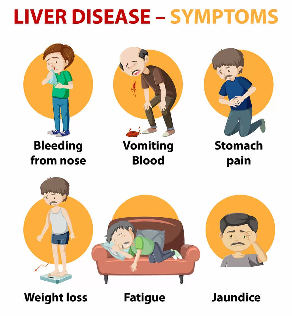
3. Systemic Symptoms
Fever and Chills:
- Infection: Fever, often accompanied by chills and rigors, may indicate an infection such as cholangitis.
Weight Loss:
- Malnutrition: Significant weight loss can occur, particularly in malignant conditions or chronic illnesses.
Anorexia and Nausea:
- Loss of Appetite: Decreased appetite and nausea are common due to the bile duct obstruction and its effects on digestion.
Malabsorption:
- Steatorrhea: Fatty, foul-smelling stools can occur due to the lack of bile in the intestines, which is essential for fat digestion and absorption.
4. Dermatological Features
Xanthomas and Xanthelasmas:
- Cholesterol Deposits: Yellowish deposits of cholesterol can appear on the skin, especially around the eyes, in chronic cholestasis.
5. Signs of Chronic Liver Disease
Spider Angiomas:
- Telangiectasias: Small, spider-like blood vessels visible on the skin, commonly seen in chronic liver disease.
Palmar Erythema:
- Red Palms: Redness of the palms, another sign of chronic liver disease.
Ascites:
- Fluid Accumulation: The buildup of fluid in the abdomen can occur in advanced liver disease or malignancy.
Coagulopathy:
- Bleeding Tendencies: Increased bleeding tendency due to impaired liver function and decreased production of clotting factors.
Unveiling the Blockage: Diagnosis of Obstructive Jaundice
Diagnosing obstructive jaundice involves a comprehensive approach that includes a detailed medical history, physical examination, laboratory tests, and various imaging techniques. The goal is to identify the cause and location of the obstruction, assess the severity of the condition, and guide appropriate treatment.
1. Medical History and Physical Examination
Medical History:
- Symptoms: Detailed description of symptoms such as jaundice (yellowing of the skin and eyes), dark urine, pale stools, itching, abdominal pain, fever, weight loss, and fatigue.
- Risk Factors: Previous history of gallstones, liver disease, pancreatic disorders, recent surgeries, family history of biliary or liver disease, alcohol consumption, and medication use.
- Duration and Onset: Understanding the timeline of symptom onset and progression.
Physical Examination:
- General Appearance: Observation of jaundice, skin scratching marks (due to itching), and signs of chronic liver disease (e.g., spider angiomas, palmar erythema).
- Abdominal Examination: Palpation for liver enlargement (hepatomegaly), tenderness, masses, and ascites (fluid in the abdomen).
- Other Signs: Signs of systemic illness, such as fever and lymphadenopathy (enlarged lymph nodes).
2. Laboratory Tests
Liver Function Tests (LFTs):
- Bilirubin: Elevated levels of total and direct (conjugated) bilirubin indicate cholestasis or bile flow obstruction.
- Alkaline Phosphatase (ALP): Elevated Alkaline Phosphatase in obstructive jaundice suggest bile duct obstruction or cholestasis.
- Gamma-Glutamyl Transferase (GGT): Elevated GGT levels often accompany elevated ALP and further suggest biliary obstruction.
- Alanine Aminotransferase (ALT) and Aspartate Aminotransferase (AST): Elevated levels indicate hepatocellular injury, which can occur alongside bile duct obstruction.
Complete Blood Count (CBC):
- White Blood Cell Count (WBC): Elevated WBCs may indicate infection or inflammation, such as cholangitis.
- Hemoglobin and Platelets: To assess for anemia or thrombocytopenia, which may occur in advanced liver disease or malignancies.
Coagulation Profile:
- Prothrombin Time (PT) and International Normalized Ratio (INR): Elevated values may indicate impaired liver function and a decreased ability to produce clotting factors.
Serum Amylase and Lipase:
- Elevated levels may indicate pancreatitis, which can cause or accompany bile duct obstruction.
3. Imaging Studies
Ultrasound:
- Initial Imaging Modality: Non-invasive, widely available, and effective for detecting gallstones, bile duct dilation, and liver abnormalities.
Computed Tomography (CT) Scan:
- Detailed Imaging: Provides detailed cross-sectional images of the abdomen, useful for identifying tumors, abscesses, and detailed anatomy of the liver, bile ducts, and pancreas.
Magnetic Resonance Imaging (MRI) and Magnetic Resonance Cholangiopancreatography (MRCP):
- High-Resolution Imaging: Non-invasive imaging that provides detailed images of the bile ducts, liver, and pancreas without the need for contrast dye. MRCP is particularly useful for visualizing bile duct obstructions.
Endoscopic Retrograde Cholangiopancreatography (ERCP):
- Diagnostic and Therapeutic Tool: Involves the use of an endoscope and contrast dye to visualize the bile and pancreatic ducts. ERCP can also be used to remove stones, place stents, and take biopsies.
Percutaneous Transhepatic Cholangiography (PTC):
- Alternative Imaging Technique: Used when ERCP is not feasible. Involves inserting a needle through the skin into the liver to inject contrast dye and visualize the bile ducts. Can also be used for therapeutic interventions like drainage.
4. Biopsy
Liver Biopsy:
- Indications: May be necessary to diagnose intrahepatic causes of obstructive jaundice, such as primary biliary cholangitis or primary sclerosing cholangitis.
Brush Cytology or Biopsy during ERCP:
- Indications: Performed to obtain tissue samples from suspected malignant strictures or tumors in the bile ducts.
Clearing the Path: Management of Obstructive Jaundice
The management of obstructive jaundice involves addressing the underlying cause of the bile duct obstruction, relieving symptoms, and preventing complications. Treatment strategies can be divided into medical management, endoscopic interventions, surgical procedures, and supportive care.
1. Medical Management
Symptomatic Relief:
- Itching (Pruritus): Managed with antihistamines, bile acid sequestrants (e.g., cholestyramine), and ursodeoxycholic acid.
- Pain: Analgesics such as acetaminophen or NSAIDs may be used for pain relief. Opioids may be required for severe pain.
Infection Control:
- Antibiotics: Administered if there is evidence of cholangitis (infection of the bile ducts) or other infections. Common antibiotics include ciprofloxacin, metronidazole, or a combination of ampicillin and gentamicin.
Nutritional Support:
- Vitamin Supplementation: Fat-soluble vitamins (A, D, E, K) may be supplemented due to malabsorption in chronic cholestasis.
- Nutritional Support: Adequate nutrition and hydration to support overall health and recovery.
2. Endoscopic Management
Endoscopic Retrograde Cholangiopancreatography (ERCP):
- Stone Removal: ERCP can be used to remove gallstones from the common bile duct.
- Stent Placement: Plastic or metal stents can be placed to bypass obstructions caused by strictures or tumors.
- Dilation: Balloon dilation can be used to widen narrowed bile ducts.
- Biopsy: Tissue samples can be taken during ERCP to diagnose malignancies or other conditions.
Endoscopic Ultrasound (EUS):
- Guided Interventions: EUS can be used to guide fine-needle aspiration of suspicious lesions and for drainage of pseudocysts or abscesses.
3. Surgical Management
Cholecystectomy:
- Gallbladder Removal: Performed in cases of gallstones causing recurrent obstruction. Can be done laparoscopically or via open surgery.
Biliary Bypass Surgery:
- Bypass Procedures: Surgical creation of a new pathway for bile flow, such as a hepaticojejunostomy or choledochojejunostomy, particularly in cases of malignancy or strictures.
Tumor Resection:
- Cancer Surgery: Surgical removal of tumors in the pancreas, bile ducts, or ampulla of Vater. This can include pancreaticoduodenectomy (Whipple procedure) for pancreatic cancer.
Liver Transplantation:
- End-Stage Liver Disease: Considered in cases of severe liver damage or biliary cirrhosis when other treatments are not effective.
4. Supportive Care
Hydration and Electrolyte Balance:
- Intravenous Fluids: To maintain hydration and correct electrolyte imbalances, especially in cases of severe vomiting or diarrhea.
Parenteral Nutrition:
- Nutritional Support: Used when oral intake is insufficient, particularly in patients with significant weight loss or malnutrition.
Monitoring and Follow-Up:
- Regular Monitoring: Regular follow-up visits to monitor liver function, bilirubin levels, and overall health status.
- Managing Complications: Ongoing management of potential complications, such as liver failure, biliary cirrhosis, and infection.
5. Oncological Treatment
Chemotherapy and Radiation:
- Adjuvant Therapy: Used for malignant causes of obstructive jaundice, such as cholangiocarcinoma or pancreatic cancer.
- Targeted Therapy: Newer treatments that target specific molecular pathways involved in cancer growth.
Palliative Care:
- Symptom Management: Focused on relieving symptoms and improving quality of life in patients with advanced malignancies.
- Support Services: Psychological support, pain management, and palliative procedures to relieve obstruction and symptoms.
Prevention of Obstructive Jaundice
Preventive measures focus on reducing risk factors and early detection:
- Healthy Diet and Lifestyle:
- Maintain a balanced diet low in saturated fats to reduce the risk of gallstones.
- Regular exercise to maintain a healthy weight.
- Avoidance of Risk Factors:
- Avoid excessive alcohol consumption.
- Manage chronic diseases like diabetes and hyperlipidemia.
- Regular Screening:
- For individuals with a family history of gallstones or biliary cancers.
- Regular check-ups and imaging for patients with chronic pancreatitis or liver diseases.
- Early Medical Intervention:
- Seek prompt medical attention for symptoms of jaundice, abdominal pain, or unexplained weight loss.
- Follow-up with healthcare providers for known biliary diseases.
Conclusion
Obstructive jaundice is a significant medical condition requiring timely diagnosis and appropriate management. Understanding the types, causes, and pathogenesis helps in choosing the right diagnostic and therapeutic approach. Preventive strategies, including a healthy lifestyle and regular medical check-ups, can reduce the risk of developing this condition. Early intervention and comprehensive care are crucial for improving outcomes and preventing complications.
FAQs
What is obstructive jaundice?
Obstructive jaundice is a condition where the flow of bile from the liver to the small intestine is blocked, causing a buildup of bilirubin in the blood. This leads to yellowing of the skin and eyes, dark urine, and pale stools.
What causes obstructive jaundice?
Common causes include gallstones, tumors (such as pancreatic or bile duct cancer), strictures (narrowing of the bile ducts), pancreatitis, and liver diseases. It can also be caused by infections and congenital anomalies.
What are the symptoms of obstructive jaundice?
Symptoms include yellowing of the skin and eyes (jaundice), dark urine, pale stools, itching, abdominal pain, nausea, vomiting, weight loss, and fever.
How is obstructive jaundice diagnosed?
Diagnosis involves a combination of medical history, physical examination, laboratory tests (liver function tests, bilirubin levels), and imaging studies such as ultrasound, CT scan, MRI, MRCP, and ERCP. A biopsy may also be required in some cases.
Can obstructive jaundice be treated?
Yes, treatment depends on the underlying cause. It may involve medical management for symptom relief, endoscopic procedures (like ERCP) to remove blockages, surgical interventions to remove tumors or bypass obstructions, and supportive care to manage complications.
What are the complications of untreated obstructive jaundice?
If left untreated, obstructive jaundice can lead to serious complications such as liver damage, infections (cholangitis), pancreatitis, biliary cirrhosis, and in severe cases, liver failure.
How can obstructive jaundice be prevented?
Prevention strategies include maintaining a healthy diet and lifestyle to reduce the risk of gallstones, managing chronic conditions like diabetes and liver disease, avoiding excessive alcohol consumption, and regular medical check-ups for early detection and treatment of biliary disorders.
Is obstructive jaundice a sign of cancer?
While obstructive jaundice can be caused by benign conditions like gallstones, it can also be a sign of malignancies such as pancreatic cancer, bile duct cancer (cholangiocarcinoma), or metastasis from other cancers. It is important to investigate the cause promptly.
What is the prognosis for someone with obstructive jaundice?
The prognosis varies depending on the cause and severity of the obstruction. Benign causes like gallstones have a good prognosis with appropriate treatment, while malignancies may require more intensive treatment and have a variable prognosis.
Can lifestyle changes help manage obstructive jaundice?
Yes, lifestyle changes such as maintaining a balanced diet, staying hydrated, avoiding alcohol, and managing underlying health conditions can support overall liver and biliary health and may help prevent recurrence of obstructive jaundice.


